3DXI

Ön most a Sonarmed Kft. régi honlapját nézegeti, ezért elképzelhető, hogy az oldal elavult információkat tartalmaz.

A legfrissebb információkért látogassa meg

új honlapunkat!


3DXI - 3D eXtended Imaging

3D XI™

3DXI - eXtended Imaging képalkotási technika

A 3D eXtended Imaging™ vagy 3D XI™ opció egy 3D képfeldolgozási eljárásokat tartalmazó csomag, mely a Multi-Slice View™, Oblique View™ és VolumeCT™ eszköztárakat tartalmazza. A Medison által ultrahang-diagnosztikai készülékekbe elsőként integrált CT és MR ekvivalens technológia segítségével az ábrázolás, a nyert klinikai adatok analízise és a diagnózis felállítása a hagyományos ultrahang-diagnosztikához képest rövidebb idő alatt, precízebben kivitelezhető.

Integration of 3D XI technologies, SONOACE is the very first in the ultrasound firm in the industry to incorporate CT and MRI-like technologies into ultrasound systems. Whether for research or in a clinical setting, Accuvix XQ with 3D XI will dramatically increase your diagnostic capabilities and clinical accuracy.


3DXI Multi Slice View - eXtended Imaging képalkotási technika

Multi-Slice View™

A Multi-Slice View™ vagy MSV™  volumen (leképezett 3D szövettömb és élő 4D megjelenítés) kiválasztott orthogonális síkja A / B / C) egymást követő, ún. sorozatmetszeteinek a képernyőn történő egyidejű ábrázolását jelenti. A tizedmilliméteres szelet-vastagság nagy pontosságú összehasonlító mérések, mérés-sorozatok végzését teszi lehetővé. A sorozatból kijelölt metszeten további képfinomítással (DMR, auto contrast, sharpen stb.) a nyert információ maximalizálható.

Multi-Slice View™ or MSV™  technology will empower you with the latest in clinical diagnostic ultrasound capabilities. Unlike diagnostic capabilities offered by existing ultrasound technologies with our new MSV you will be able to view and diagnose clinical cases much faster and most importantly with more precision and accuracy than ever before. Whether in a clinical setting or for research, Multi-Slice View available in Accuvix XQ ultrasound system will take your diagnostic capabilities to new height of clinical accuracy and will become an essential solution to all your imaging needs.   
 

3DXI Oblique View ultrahang képalkotási technika

Oblique View™

Az Oblique View™  a referenciasík választott pontja (ROI) térbeli összefüggéseinek vizsgálatára szolgál. Adott forgatási pont körüli metszetek megjelenítése (static line), tetszőlegesen választott forgatási pont körüli metszetek megjelenítése (dynamic line) és a referencia síkon kijelölt kontúr révén nyert görbült metszet egy síkban való megjelenítése (contour ill. „medvebőr” ábrázolás).

Oblique View™ is imaging technology which enables you to examine and view 3D volume data in various planes without limitations. You are able to select the exact portion of the 3D data that you would like to examine thus allowing for more complete visual examination and better understanding of the correlation between organs and other organs or areas within the region of interest.   
 

3DXI Volume CT ultrahang képalkotási technika

VolumeCT View™

A VolumeCT View™ technológia Cross Volume CT funkciója a tér 3 orthogonális síkjának egy metszéspontban való egyidejű megjelenítését, elforgatását, nagyítását, referenciasíkkal történő áttekintését - felszeletelését, analízisét teszi lehetővé, míg a  Cube Sectional View funkció egy virtuális kocka oldalaira vetítve látja el ugyanezt a feladatot. A VolumeCT View™ a háromdimenziós orientáció megkönnyítése mellett  a térbeli összefüggések pontosabb ábrázolásának eszköze.

VolumeCT View™ technology with Cross View and Cube Sectional View functions enables you to perform multiple examinations on multiple regions of interest and visually displays their relationships from data obtained with just one 3D volume scan. Therefore, eliminating the need for multiple scans and making it possible to examine the data even after the initial scan session.   


Multi-Slice View™or MSV™ technology will empower you with the latest in clinical diagnostic ultrasound capabilities. Unlike diagnostic capabilities offered by existing ultrasound technologies with our new MSV you will be able to view and diagnose clinical cases much faster and most importantly with more precision and accuracy than ever before. Whether in a clinical setting or for research, Multi-Slice View available in Accuvix XQ ultrasound system will take your diagnostic capabilities to new height of clinical accuracy and will become an essential solution to all your imaging needs.
 

Nuchal Translucency


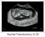
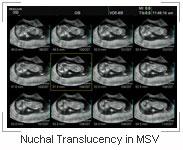
In diagnosing for Nuchal Translucency, MSV Technology really shines. As seen here, compared to the limited measuring capabilities of 2D image, our MSV is able to display the entire range from the maximum and minimum measurements, giving you a more precise assessment. In this case of measuring for NT in 12 week fetus, the diameter measurement was shown to be 3.6mm using the 2D measuring method. Where as utilizing MSV, the NT measurements in the same case ranged from a maximum of 4.6mm to minimum of 3.5mm thus giving you an more accurate diagnosis. Additionally, the MSV measurement diagnoses was accomplished by scanning the patient just once.
 

Nuchal Translucency in 2D Nuchal Translucency in MSV


 Study of Fetal Brain


MSV can also be utilized in the very important area of assessing infant development. In these case studies, MSV was used to assess the development of two fetuses in their pivotal early gestation stages. With MSV, it is possible to examine the complete section of the fetal brain for nuchal transluency in axial, CSP, lateral ventricle and cellebelium developments.

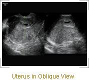
In this case study of the abnormal uterus (bicoronate uterus), you can clearly see the clinical advantages of MSV technology. In the 2D sagital plane view of bicornate uterus, it is very difficult to distinguish the coronate, let alone diagnose it accurately. But with MSV image on the right, you can clearly see and track the coronal in c-plane view and thanks to the sequential imaging capabilities, you can also clearly see the progression of the coronal throughout the uterus. In addition to easily being able to track the difference, you can also view and measure the difference in angles throughout the entire range of the uterus, which is not possible with current ultrasound technologies.


Oblique View™ is imaging technology which enables you to examine and view 3D volume data in various planes without limitations. You are able to select the exact portion of the 3D data that you would like to examine thus allowing for more complete visual examination and better understanding of the correlation between organs and other organs or areas within the region of interest.
 

 Fetal Ascites


Fetal Ascites in Static Line Oblique View Fetal Ascites in Multi-Planar Imaging By applying the Contour Oblique View function to the irregular contours of this normal 2D image of bicornate uterus (left inset) from the original 3D scan data set, it is possible to easily obtain an cross sectional image of the uterus from the fundus to the cervix (right inset).

Other images from this 3D data set can also explored for a more thorough examination, this type of versatility was not available in the past without performing multiple scanning sessions. But with 3D-XI, it is now possible to accomplish multiple examinations with data obtained from just one simple 3D scan.


 Bicornate Uterus


By applying the Contour Oblique View function to the irregular contours of this normal 2D image of bicornate uterus (left inset) from the original 3D scan data set, it is possible to easily obtain an cross sectional image of the uterus from the fundus to the cervix (right inset). Other images from this 3D data set can also explored for a more thorough examination, this type of versatility was not available in the past without performing multiple scanning sessions. But with 3D-XI, it is now possible to accomplish multiple examinations with data obtained from just one simple 3D scan.


VolumeCT View™ technology with Cross View and Cube Sectional View functions enables you to perform multiple examinations on multiple regions of interest and visually displays their relationships from data obtained with just one 3D volume scan. Therefore, eliminating the need for multiple scans and making it possible to examine the data even after the initial scan session.
 

 Fetal Abdomen


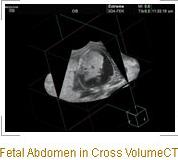
In this case involving a seven month fetus examination, by utilizing VolumeCT and its Cross View function, it is feasible to examine nearly all of the organs and its relationship within the entire abdomen in one setting and with just one simple 3D scan. By manipulating the X, Y and Z axes of the Cross View function of VolumeCT, the kidney is shown on the sagital plane and stomach in the coronal plane while the bladder is displayed on the transverse plane, this type of complete 3D imaging allows for better understanding of the correlation between the all three organs as well as other areas within the abdomen.


 Fetal Ascites


The image on the right illustrates another function of the VolumeCT, the Cube Sectional View. Where the Cross View of VolumeCT looks at the three axes of the image data from the internal to the exterior of the data set and its relationships contained within, the Cube Sectional View is able to examine the 3D data from exterior towards interior within the data set. In this clinical case, from a regular 3D data set with application of Cube Sectional View, all sides of the volume data can be examined in a "3D cube like" image with the entire six sides showing the images from the data set. Therefore, it is possible to view the entire ascites and also the enlarged stomach of this fetus on one side of the cube while and the caudal of the liver is shown on the opposite side.


3D XI PC Viewer 60 napos próbaverzió számítógépre letöltése


A tulajdonsággal rendelkező ultrahang készülékeink:

(a lista már nem teljes, mivel ez a honlap már nem tartalmazza a 2019 óta megjelent legújabb készülékeinket)

Régi honlap - elavult információk

Ön most a Sonarmed Kft. régi honlapját nézegeti, ezért elképzelhető, hogy az oldal elavult információkat tartalmaz.

A legfrissebb információkért látogassa meg

új honlapunkat!

Új Samsung ultrahang készülékek

Új Samsung ultrahang készüléket keres?

Jó helyen jár!

Kattintson a logóra!

Samsung ultrahang készülékek minden orvosi területre

Samsung WS80A Elite